
What Lumbar Spinal Stenosis Means for Your Mobility
Dealing with this in San Diego? Same-day appointments are often available at our 4 clinics.
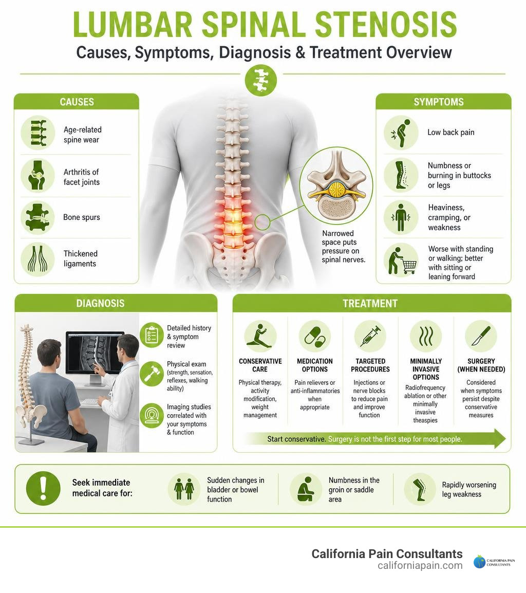
Lumbar spinal stenosis is a narrowing of spaces in the lower back that can squeeze spinal nerves. It may cause low back pain, numbness, burning pain, heaviness, cramping, or weakness in the buttocks and legs – especially when standing or walking. Symptoms often improve when you sit down or lean forward, such as over a shopping cart.
The most common cause is age-related spine wear, including disc changes, arthritis in the facet joints, bone spurs, and thickened ligaments. But imaging alone does not tell the whole story: many people have spinal narrowing without symptoms. A proper evaluation connects scan findings with your pain pattern, strength, sensation, walking ability, and overall function.
The good news is that surgery is not the first step for most people. Physical therapy, activity changes, medication when appropriate, and targeted procedures may help improve comfort and walking tolerance. Sudden bladder or bowel changes, numbness in the groin or saddle area, or rapidly worsening leg weakness need emergency medical care.
I am Dr. Zach Cohen, a double board-certified anesthesiologist and chronic pain physician with training in interventional spine care and minimally invasive procedures. In treating Lumbar spinal stenosis and other chronic pain conditions, I focus on clear diagnosis and personalized options that support movement without relying only on medication.
Basic Lumbar spinal stenosis vocab:
Understanding Lumbar Spinal Stenosis: Anatomy and Types
To understand why lumbar spinal stenosis causes walking discomfort and aching legs, it helps to look at the underlying architecture of your lower back. The lumbar spine consists of five robust vertebrae (labeled L1 through L5) that sit stacked atop one another, connecting your upper torso to your pelvis. These bony segments shield a central passageway called the spinal canal.
While the solid spinal cord ends around the upper lumbar spine (specifically L1–L2), it transitions into a delicate spray of nerve roots known as the cauda equina (Latin for “horse’s tail”). These nerves float inside a protective, fluid-filled sheath called the thecal sac before branching out through small bony doorways to power your hips, thighs, calves, and feet.
As detailed in the Johns Hopkins Medicine overview on spinal canal narrowing, when the protective space inside this bony canal constricts, mechanical friction and localized blood flow restriction put pressure on the passing nerve pathways.
Central, Lateral Recess, and Foraminal Stenosis
Spinal canal narrowing is not a one-size-fits-all condition; it occurs in distinct anatomical zones within the vertebra:
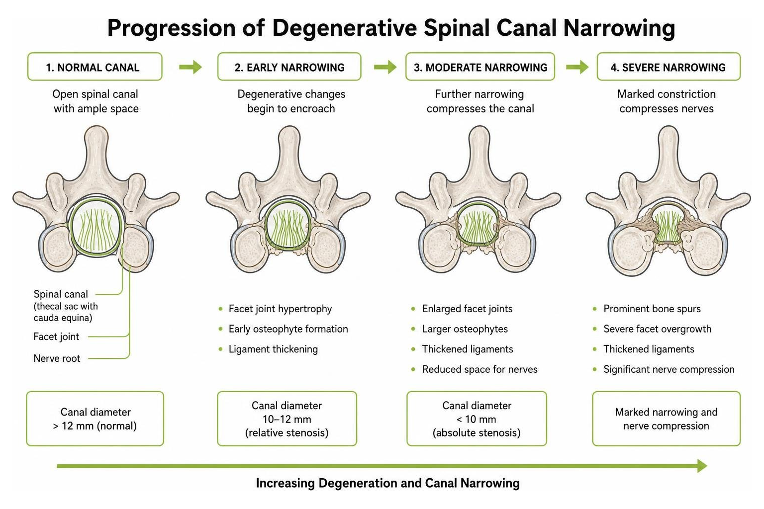
- Central Canal Stenosis: The primary hollow tube running through the center of the vertebra narrows, squeezing the thecal sac and cauda equina. Clinically, a normal canal measures over 12 mm in midsagittal diameter; measurements between 10 and 12 mm indicate relative stenosis, while an opening under 10 mm represents absolute central stenosis.
- Lateral Recess Stenosis: Also known as the “subarticular zone,” this is the internal corridor a nerve root navigates just before exiting the main canal. Hypertrophic facet joints often pinch nerves here.
- Neural Foraminal Stenosis: The lateral exit portals (the neuroforamina) where nerve roots leave the spine to travel down your legs become constricted, frequently due to collapsing disc space and osteophyte formation.
Congenital vs. Acquired Degenerative Stenosis
Most adults develop acquired degenerative stenosis, a progressive narrowing that typically surfaces after age 50 as a result of long-term mechanical wear. Up to 95% of people exhibit some degree of degenerative spinal change on imaging by their fifth decade.
Conversely, some individuals are born with congenital spinal stenosis. These patients possess anatomically short pedicles—the bony pillars framing the spinal canal—creating an inherently narrower passageway from birth. Although congenital stenosis is less common and occurs more often in men, symptoms can emerge much earlier in life, frequently between ages 30 and 50, when mild everyday wear further tightens an already crowded canal.
Key Causes and Risk Factors of Spinal Canal Narrowing
Understanding the underlying back pain causes helps clarify why conservative, targeted treatments are so effective at addressing symptoms.
Primary Underlying Causes of Lumbar Spinal Stenosis
Spinal narrowing stems from a predictable sequence of joint and tissue changes in the lower back:
- Osteoarthritis and Facet Arthropathy: As cartilage inside the stabilizing facet joints wears thin, friction triggers local inflammation. The body responds by generating excess bone spurs (osteophytes) that protrude inward toward spinal nerves.
- Intervertebral Disc Desiccation and Bulging: Healthy spinal discs consist largely of water. Over time, these shock absorbers lose hydration, collapse in height, and bulge outward into the canal space.
- Ligamentum Flavum Hypertrophy: The flexible yellow ligament lining the rear wall of the spinal canal thickens and loses elasticity over time, buckling forward into the dural sac.
- Degenerative Spondylolisthesis: When supporting discs and facet joints weaken, one lumbar vertebra can slip forward over the one beneath it, creating an internal shelf that abruptly narrows the neural canal, as highlighted in the MSD Manual guide to lumbar stenosis.
Secondary Risk Factors and Degenerative Changes
Beyond natural mechanical wear, several contributing risk factors accelerate canal narrowing:
- Age: Symptomatic stenosis rises sharply with age, documented in 1.9% of adults in their 40s compared to nearly 11% of adults in their 70s.
- Repetitive Postural Load: Decades of heavy physical labor or prolonged upright compression place elevated shear stress on the lumbar facet joints.
- Skeletal Trauma and Prior Surgeries: Fractures or post-surgical scar tissue can alter internal spinal mechanics and reduce canal volume.
- Tobacco Use: Nicotine constricts microvascular blood supply to intervertebral discs, accelerating disc collapse and facet degeneration.
Recognizing the Symptoms and Warning Signs
Lumbar spinal stenosis symptoms often develop gradually rather than appearing overnight. Patients commonly describe dull, aching axial low back pain alongside deep gluteal tightness, calf heaviness, tingling sensations, and radiating leg discomfort.
Neurogenic Claudication and the Shopping Cart Sign
The hallmark clinical presentation of lumbar spinal stenosis is neurogenic claudication (sometimes called pseudoclaudication). Unlike typical muscle soreness, neurogenic claudication is strictly posture-dependent:
- Aggravating Postures: Standing tall, walking down flat hallways, or stepping downhill forces the lumbar spine into extension (backward arching). Extension increases lumbar lordosis, causing the ligamentum flavum to buckle forward and bone spurs to pinch nerves, triggering burning pain, cramping, and heavy legs.
- Relieving Postures: Sitting down or bending forward flexes the spine, flattening lumbar lordosis and widening the spinal canal diameter.
This posture dynamic gives rise to the classic “shopping cart sign.” Patients who struggle to walk 50 yards across a parking lot can often stroll through a grocery store without leg discomfort simply by leaning forward over the handle of a shopping cart. For the same anatomical reason, riding a stationary bicycle or walking uphill feels far more manageable than walking downhill.
Red Flag Emergency: Cauda Equina Syndrome
While most spinal stenosis cases progress slowly and can be managed conservatively, profound compression of the lower nerve roots can trigger Cauda Equina Syndrome. This is a true surgical emergency.
Seek immediate emergency medical evaluation if you develop:
- Sudden loss of bowel or bladder control (incontinence or urinary retention)
- “Saddle anesthesia” (loss of sensation around the groin, buttocks, perineum, and inner thighs)
- Rapidly progressive motor weakness, such as severe bilateral foot drop (inability to lift the front of your foot)
Accurate Diagnosis and Differential Evaluation
An accurate clinical assessment is critical because imaging abnormalities alone do not always correlate with daily symptoms. In fact, radiological studies demonstrate that up to 80% of individuals over age 60 exhibit moderate-to-severe canal narrowing on imaging while remaining completely pain-free. Establishing an accurate back pain diagnosis requires connecting your scan results with real-world functional tests.
During a comprehensive physical examination, clinicians perform targeted assessments:
- Kemp’s Provocative Test: Extending and rotating the lumbar spine to reproduce canal narrowing and assess radicular pain.
- Five-Repetition Sit-to-Stand (5R-STS): Assessing lower-body motor power and functional endurance.
- Romberg and Gait Testing: Checking for wide-based walking patterns or sensory balance deficits.
Clinical Examination and Neurogenic vs. Vascular Claudication
One of the most important diagnostic distinctions is separating neurogenic claudication (nerve compression in the spine) from vascular claudication (inadequate blood flow caused by peripheral artery disease). Both conditions cause leg pain during walking, but their triggers and relief mechanisms differ substantially.
| Clinical Feature | Neurogenic Claudication (Spinal Stenosis) | Vascular Claudication (Circulatory Insufficiency) |
|---|---|---|
| Primary Cause | Nerve compression & ischemia in the spinal canal | Arterial insufficiency in lower extremities |
| Pain Characteristics | Dull ache, burning, heaviness, weakness, numbness | Tight cramping, squeezing muscle pain |
| Pain Distribution | Buttocks, thighs, calves; often bilateral | Calves, thighs, or arches; muscular focus |
| Postural Sensitivity | Relieved by forward flexion (bending over, sitting) | Posture-independent; relieved by resting muscles |
| Response to Standing Still | Pain persists while standing upright | Pain resolves quickly within minutes of stopping |
| Uphill vs. Downhill Walking | Uphill is easier (spine flexes forward) | Uphill is more painful (muscles demand more oxygen) |
| Pedal Pulses | Normal and readily palpable | Diminished, asymmetric, or absent |
| Skin Temperature & Color | Normal lower extremity skin changes | Pallor on elevation, skin coolness, hair loss |
Advanced Diagnostic Imaging and Nerve Studies
When physical signs suggest significant stenosis, advanced diagnostic tools help map neural compression:
- Magnetic Resonance Imaging (MRI): The gold-standard diagnostic tool. MRI clearly depicts soft tissue structures, disc herniations, facet joint overgrowth, and the nerve root sedimentation sign (a key radiologic indicator where compressed nerve roots fail to settle along the back wall of the dural sac on supine cross-sections).
- Computed Tomography (CT) & CT Myelography: When patients have implanted pacemakers or metal hardware preventing MRI use, CT myelography utilizes radiopaque contrast dye to provide sharp cross-sectional views of thecal sac constriction.
- Flexion-Extension Radiographs: Weight-bearing dynamic X-rays evaluate whether vertebrae slip abnormally during movement, ruling out dynamic spondylolisthesis.
- Electromyography (EMG) and Nerve Conduction Studies: Electrodiagnostic testing verifies whether nerve root impingement is causing physiological conduction delays down the leg, as highlighted in the StatPearls clinical review on lumbar spinal stenosis.
Comprehensive Management: From Non-Surgical Care to Surgery
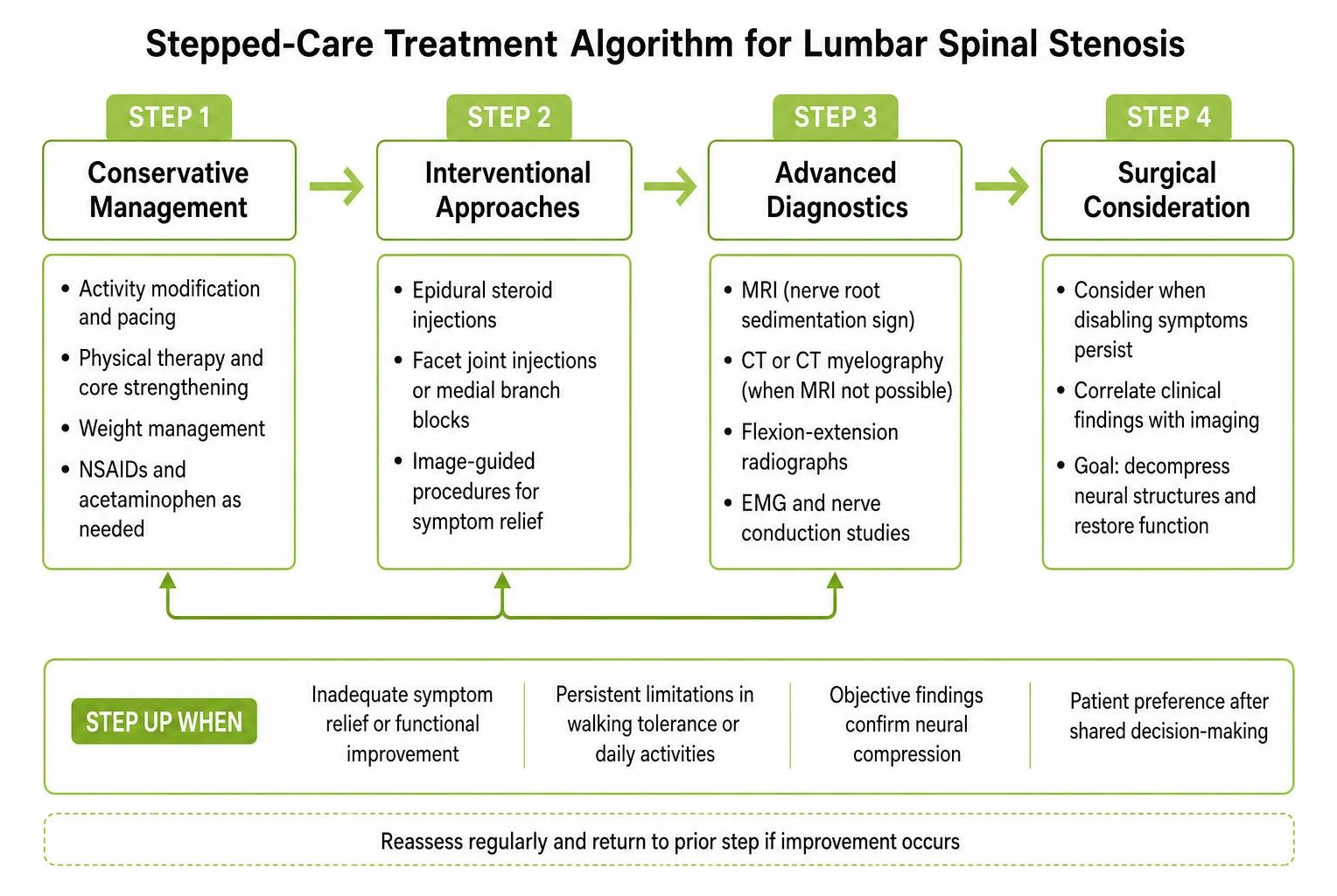
Managing spinal stenosis effectively relies on a structured, stepped-care pathway. Because the natural history of mild-to-moderate stenosis remains stable in many individuals, conservative and interventional approaches serve as the primary standard of care. At California Pain Consultants, our multidisciplinary clinics throughout San Diego, Kearny Mesa, Chula Vista, Rancho Bernardo, La Mesa, and Miramar focus on restoring walking tolerance and functional independence without relying on heavy daily medications or immediate open spine surgery.
Our practical approach is detailed extensively in our non-surgical pain relief guide.
Evidence-Based Non-Surgical Treatments for Lumbar Spinal Stenosis
Initial management focuses on reducing mechanical pressure, restoring core stability, and controlling localized inflammation:
- Targeted Physical Therapy: Structured pain management physical therapy prioritizes a flexion-biased protocol. Therapists guide patients through tailored exercises for lower back pain that strengthen the transversus abdominis, gluteals, and pelvic floor.
- Therapeutic Stretching: Performing dedicated lower back stretches—such as gentle knee-to-chest maneuvers and seated pelvic tilts—opens the neuroforamina and decompresses tense lumbar musculature.
- Non-Steroidal Anti-Inflammatory Drugs (NSAIDs): Short courses of oral anti-inflammatories help reduce peri-neural swelling around irritated nerve roots.
- Neuromodulatory Medications: When nerve pain or burning radiculopathy persists, targeted nerve pain medication (such as gabapentinoids) helps quiet hypersensitive peripheral nerve signaling.
Interventional Procedures and Targeted Injections
When oral medications and physical therapy alone do not fully resolve walking limitations, advanced interventional pain management offers precise, image-guided relief:
- Epidural Steroid Injections: Utilizing live fluoroscopic guidance, clinicians deliver targeted anti-inflammatory solutions into the interlaminar or transforaminal space, achieving lasting epidural back pain relief by washing away inflammatory cytokines surrounding compressed nerves.
- Facet Joint Injections and Diagnostic Blocks: Precise fluoroscopic back pain injections soothe irritated facet joint capsules that contribute to localized axial lower back aching.
- Radiofrequency Ablation (RFA): For patients suffering from severe concurrent facet arthritis, radiofrequency ablation therapy uses gentle thermal energy to disrupt pain signals from sensory medial branch nerves, providing 9 to 18 months of targeted relief.
- Regenerative Medicine: Emerging regenerative injection therapy protocols aim to improve tissue resilience and joint stability around chronic degenerative spinal segments.
- Spinal Cord Stimulation (SCS): In complex or treatment-resistant cases, advanced spinal cord stimulator therapy uses mild electrical pulses along the dorsal columns to replace persistent leg aching with soothing sensations.
When Is Surgical Decompression Recommended?
Approximately 20% to 40% of individuals with moderate-to-severe lumbar stenosis eventually require surgical evaluation within a decade of diagnosis. Surgery is indicated when severe pain, progressive muscle weakness, or restricted walking tolerance significantly impairs daily quality of life despite 3 to 6 months of comprehensive non-surgical management.
Surgical options include:
- Minimally Invasive Decompression: Procedures such as the MILD (Minimally Invasive Lumbar Decompression) technique remove small portions of thickened ligamentum flavum through an incision smaller than a dime, preserving bony architecture.
- Laminectomy / Laminotomy: Open or tubular decompression where the surgeon removes the rear lamina bone and overgrowing spurs to permanently widen the central canal.
- Spinal Fusion: If spinal stenosis is accompanied by dynamic instability, such as a high-grade spondylolisthesis, surgeons may combine decompression with minimally invasive spine surgery and instrumentation to stabilize shifting vertebrae.
When evaluating your options, consulting spine specialists ensures that procedural choices match your specific anatomical pathology and functional goals.
Frequently Asked Questions About Lumbar Spinal Stenosis
How do you know if your back pain is caused by lumbar spinal stenosis?
The defining characteristic of lumbar spinal stenosis is posture-dependent leg pain known as neurogenic claudication. If your back, buttock, or calf pain worsens after standing upright or walking short distances, but eases when you sit down or lean forward over a counter or shopping cart, spinal canal narrowing is a likely contributor. A clinical exam paired with an MRI confirms the diagnosis.
Can lumbar spinal stenosis resolve on its own without surgery?
While the underlying bony changes (such as facet spurs or thickened ligaments) do not disappear on their own, the symptoms of spinal stenosis frequently stabilize or improve without surgery. Clinical studies show that up to 50% of patients experience significant pain relief and improved walking ability through non-surgical care, including physical therapy, core conditioning, anti-inflammatory treatments, and targeted fluoroscopic injections.
What daily lifestyle modifications help manage spinal stenosis symptoms?
Practical adjustments make a measurable difference in daily comfort:
- Adopt a Flexion-Biased Routine: Choose stationary bicycling or water aerobics over steep downhill hiking or high-impact running.
- Ergonomic Posture Adjustments: Use footrests while sitting to keep hips slightly flexed, and sleep on your side with a pillow between your knees.
- Maintain Core Strength and Healthy Weight: Reducing mechanical strain on the lumbar spine takes pressure off the facet joints and supporting ligaments.
- Pace Your Walking: Take short sitting breaks before severe claudication pain sets in, gradually extending your total walking distance over time.
Conclusion: Restoring Mobility and Finding Lasting Relief
Lumbar spinal stenosis can limit your ability to walk, travel, and enjoy everyday activities, but it does not have to dictate your mobility. With an accurate clinical diagnosis, a clear understanding of your symptom triggers, and a modern stepped-care treatment plan, significant and lasting relief is achievable.
At California Pain Consultants, our double board-certified pain specialists provide compassionate, individualized care across our convenient locations in San Diego, Kearny Mesa, Chula Vista, Rancho Bernardo, La Mesa, and Miramar. If you or a family member are struggling with back stiffness, leg cramping, or reduced walking tolerance, explore our comprehensive treatment for chronic back pain to schedule your evaluation and take the first step toward restoring your active lifestyle.