
Understanding How Your Low Back Muscles Protect and Move Your Spine
Dealing with this in San Diego? Same-day appointments are often available at our 4 clinics.
Your low back muscle system serves as an internal brace and engine for your entire upper body. It carries out three essential jobs:
- Segmental stability: Deep local stabilizers (like the multifidus) lock individual spinal joints in place to keep them aligned.
- Dynamic movement: Large superficial muscles (like the erector spinae) generate the power needed to stand up, bend, twist, and lift.
- Load distribution: Balanced spinal muscles absorb daily physical forces, shielding your spinal discs and nerves from excessive pressure.
When these muscles become weak, fatigued, or uncoordinated, extra stress falls directly onto spinal ligaments and joints, frequently leading to pain and protective muscle spasms.
Nearly 80% of adults experience lower back pain during their lives. Fortunately, most episodes stem from treatable muscular imbalances rather than permanent spinal damage.
I am Dr. Zach Cohen, a double board-certified anesthesiologist and pain management specialist with fellowship training from the University of California San Diego. Over my career, I have guided thousands of patients toward lasting relief by restoring healthy low back muscle function through personalized, non-surgical treatment plans.
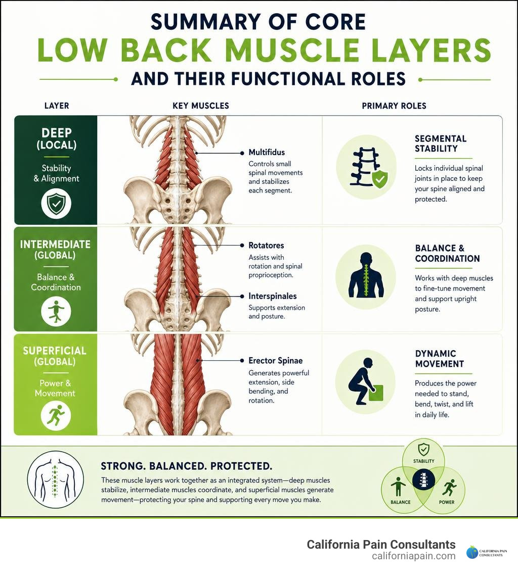
Terms related to low back muscle:
Primary Low Back Muscle Groups and Their Functional Roles
To understand spinal health, we must view the lumbar spine as an architectural suspension bridge. The bones and discs provide the vertical pillars, but the muscles and ligaments serve as the dynamic cables holding everything upright against gravity.
When analyzing back muscular anatomy, we divide the musculature into two functional categories: deep local stabilizers and superficial global mobilizers.
| Functional Category | Primary Muscles Included | Anatomical Depth | Primary Biomechanical Role | Predominant Muscle Fiber Type |
|---|---|---|---|---|
| Deep Local Stabilizers | Lumbar Multifidus, Rotatores, Interspinales | Deep layer (adjacent to vertebrae) | Segmental stiffness, micro-motion control, joint position sensing (proprioception) | Type I (Slow-Twitch, Fatigue-Resistant) |
| Superficial Global Mobilizers | Longissimus Thoracis, Iliocostalis Lumborum, Quadratus Lumborum | Intermediate to superficial layers | Torque generation, gross spinal movement (extension, lateral tilt), load distribution | Mixed / Type II (Fast-Twitch, Power-Generating) |
Both layers must work in precise coordination. If you’d like an extensive breakdown of the broader muscular network across the spine, explore our comprehensive guide to back muscles.
Deep Stabilizers: The Lumbar Multifidus and Rotatores
The lumbar multifidus is the cornerstone of active segmental stability in the lumbar spine. Tightly packed along the posterior vertebral arches, these short, powerful muscle fascicles cross between two and four spinal segments.
According to a detailed scoping review on lumbar multifidus morphology, the multifidus accounts for over two-thirds of active segmental stiffness in the lumbar “neutral zone”—the small range of physiological movement where spinal ligaments remain slack. Because the deep multifidus possesses short lever arms, it does not create massive sweeping trunk movements; instead, it generates immediate compressive force across intervertebral joints, preventing abnormal shearing and micro-slippage during daily activity.
Lying immediately beneath and alongside the multifidus are the rotatores (brevis and longus) and interspinales. Richly innervated with muscle spindles, these deep muscular structures act as sophisticated sensory proprioceptors, telling your brain the exact position of your vertebrae in space.
Global Mobilizers: Erector Spinae and Quadratus Lumborum
Positioned further away from the spinal axis are the global mobilizing muscles, designed with long lever arms to produce powerful torque:
- Erector Spinae: Comprising the longissimus thoracis and iliocostalis lumborum, these massive paired columns run alongside the spine from the sacrum and iliac crest up into the ribs and thoracic vertebrae. They provide the primary extensor force that allows you to lift heavy objects and maintain an upright posture.
- Quadratus Lumborum (QL): Located in the posterior abdominal wall lateral to the lumbar vertebrae, the QL connects the 12th rib, the lumbar transverse processes, and the iliac crest. As detailed in the clinical anatomy of the quadratus lumborum, the QL generates about 10 N of extension force compared to 100 N from the erector spinae and 150 N from the multifidus. Rather than acting as a prime mover, it serves as a dynamic myofascial tension pivot linking the ribcage, pelvis, and lumbar spine.
How Spinal Muscles Maintain Biomechanical Stability and Movement
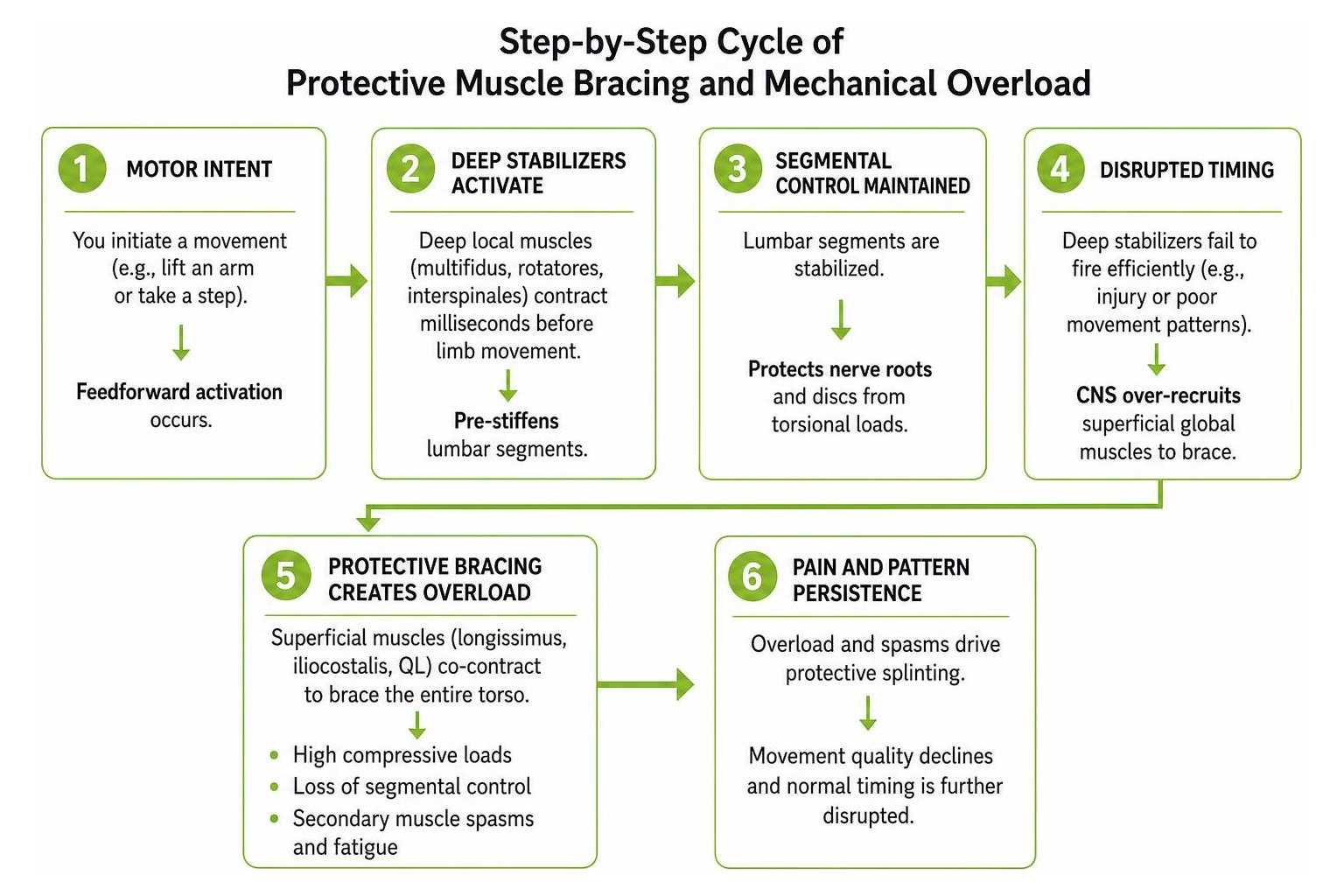
Spinal movement is not simply about brute muscular strength; it depends on exquisite neuromuscular timing. When you decide to lift an arm or take a step, your central nervous system utilizes a strategy known as feedforward activation or anticipatory postural adjustment.
In a healthy system, the deep local back muscles contract milliseconds before your limbs move. This anticipatory contraction pre-stiffens the lumbar segments, protecting sensitive nerve roots and discs from unpredictable torsional loads.
Segmental Control vs. Global Motion
When healthy sensorimotor timing is disrupted—often due to an acute injury or poor movement patterns—the deep local stabilizers fail to fire efficiently. The central nervous system compensates by over-recruiting superficial global mobilizers like the longissimus and iliocostalis to brace the entire torso.
While this protective bracing prevents immediate spinal collapse, it creates a serious mechanical tradeoff:
- High Compressive Loads: Superficial muscle co-contraction significantly increases intervertebral disc compression.
- Loss of Segmental Control: Global muscles cannot control individual intervertebral shear, leaving single motion segments vulnerable to micro-instability.
- Secondary Muscle Spasms: Overworked superficial muscles quickly exhaust their metabolic reserves, resulting in painful cramps and protective splinting.
Understanding this protective mechanism is vital when treating a pulled muscle in back tissues, where localized reflex arcs cause widespread muscle tightness far beyond the initial site of strain.
The Interconnected Kinetic Chain: Gluteal and Abdominal Synergy
The lumbar musculature does not function in isolation. It relies on coordinated synergy across the entire lumbopelvic-hip complex:
- Gluteus Medius and Maximus: Strong gluteal muscles drive hip extension and pelvis stabilization during walking. When the gluteus medius is weak or inhibited (often from prolonged sitting), the quadratus lumborum overcompensates by hiking the hip during each stride. This compensatory pattern frequently manifests as a combined pulled muscle lower back and buttock presentation.
- Transversus Abdominis and Psoas Major: The deep transversus abdominis wraps around the torso like an internal corset, tensioning the thoracolumbar fascia in tandem with the multifidus. Meanwhile, a balanced psoas major stabilizes the front of the lumbar spine without pulling the pelvis into excessive anterior tilt (hyperlordosis).
- Hamstring Tension: Chronically tight hamstrings restrict normal anterior pelvic tilt during bending, forcing the lumbar spine to flex excessively and overloading the paraspinal extensor muscles.
Structural and Neuromuscular Changes in Acute vs. Chronic Back Pain
When lower back pain transitions from an acute strain (lasting under 4 weeks) into chronic low back pain (persisting past 12 weeks), distinct structural and neurological adaptations occur within the back muscles.
In our clinical practice across San Diego, La Mesa, and Chula Vista, we regularly observe these structural adaptations on diagnostic imaging:
Fatty Infiltration and Atrophy in Low Back Muscle Tissues
In chronic back pain, muscle changes go far beyond simple disuse. Published research on paraspinal muscle quality and activation demonstrates that paraspinal muscle health is dictated primarily by functional cross-sectional area (FCSA) rather than raw muscle volume.
- Intramuscular Adipose Tissue (IMAT): In chronic conditions, non-contractile fat cells infiltrate between muscle fascicles. This muscle-to-fat infiltration index rises markedly at the L4-L5 and L5-S1 levels.
- Fiber Type Transformation: High-endurance, fatigue-resistant slow-twitch (Type I) muscle fibers undergo phenotypic shifts into fatigable fast-twitch (Type II) fibers.
- Proinflammatory Cytokines: Following disc injury or persistent mechanical irritation, localized expression of inflammatory cytokines like TNF and IL-1β drives M1 macrophage infiltration, promoting fibrotic scarring within deep multifidus tissue.
Furthermore, a study on lumbar muscle morphology in segmental instability showed that while deep multifidus atrophy occurs universally in chronic back pain, patients with segmental instability often exhibit compensatory hypertrophy of the superficial erector spinae at L4-L5 as the body attempts to mechanically stabilize the spine.
Sensorimotor Smudging and Altered Activation Patterns in Low Back Muscle Function
Chronic pain fundamentally alters how your brain communicates with your spinal musculature. Corticomotor mapping reveals that the distinct brain representations for the deep multifidus and superficial erector spinae begin to overlap—a phenomenon known as cortical smudging.
This neurological reorganization leads to measurable functional deficits:
- Spatial Activity Redistribution Failure: High-density surface electromyography (EMG) shows that healthy individuals shift muscular activity dynamically across different paraspinal muscle zones as they tire. Patients with chronic pain lose this adaptive shifting, overloading localized cranial muscle bands.
- Diminished Endurance Capacity: On the standardized Biering-Sorensen back endurance test, healthy pain-free individuals sustain holds for 78 to 221 seconds, whereas chronic back pain patients fatigue in just 39 to 144 seconds.
- Reflexive Guarding: Persistent pain creates hyperactive motor reflexes. If you are struggling with severe acute muscle guarding, reviewing how to relieve lower back spasms safely in 5 simple steps can help calm acute neuromuscular tension.
Evidence-Based Rehabilitation Strategies to Restore Muscle Health
Rehabilitating the lumbar musculature requires an individualized, step-by-step approach rather than generic core routines. At California Pain Consultants, our board-certified physicians work with patients throughout San Diego County to restore functional capacity using evidence-based conservative pathways.
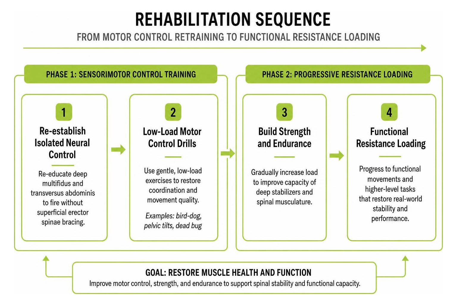
Motor Control Training vs. Progressive Resistance Loading
Restoring compromised back muscles involves two coordinated phases:
- Phase 1: Sensorimotor Control Training: Before adding heavy resistance, you must re-establish isolated neural control of the deep multifidus and transversus abdominis. Gentle, low-load motor control drills (such as quadruped bird-dogs, pelvic tilts, and dead bugs) re-educate the brain to fire stabilizing muscles without triggering superficial erector spinae bracing.
- Phase 2: Progressive Resistance Loading: Once proper recruitment patterns are established, we transition patients to progressive resistance exercises to stimulate lean muscle hypertrophy, reverse fatty infiltration, and build fatigue endurance.
For structured routines that safely incorporate these principles, see our comprehensive guide on exercises for lower back pain as well as our targeted library of exercises for low back pain relief.
Pathology-Specific Stretching and Mobility Protocols
Not all back pain benefits from the same stretching routine. Exercises must match your specific underlying spinal pathology:
- Discogenic Pain (Extension Bias): If pain stems from an irritated intervertebral disc that worsens with sitting or forward bending, gentle standing lumbar extensions and prone press-ups (McKenzie protocols) help centralize symptoms and relieve posterior disc pressure.
- Spinal Stenosis or Facet Joint Irritation (Flexion Bias): If discomfort increases with backward bending and standing, flexion-based stretches like single knee-to-chest and seated forward bends open the intervertebral foramina and ease facet joint loading.
- Kinetic Chain Mobility: Restoring length in tight hamstrings, hip flexors, and the piriformis relieves secondary pulling forces on the pelvis.
To safely integrate flexibility routines into your day, consult our detailed lower back stretches guide and learn how to stretch lower back tissues without triggering nerve flare-ups.
Frequently Asked Questions About Low Back Muscles
Why does bed rest worsen low back muscle pain?
Strict bed rest is one of the most counterproductive approaches for lower back muscle recovery. Complete immobilization triggers rapid paraspinal disuse atrophy within days, reduces blood circulation, and leads to connective tissue stiffness. Furthermore, bed rest reinforces fear-avoidance behaviors and neural inhibition. Maintaining gentle, tolerated daily walking preserves muscular tone and accelerates tissue healing.
How does multifidus atrophy differ between acute and chronic pain?
In acute low back pain, multifidus shrinkage is driven primarily by neural reflex inhibition—a localized spinal reflex where pain signals actively shut down muscle firing without immediate structural cell death. In chronic low back pain, persistent non-use and localized inflammatory cytokine activity cause true structural remodeling, replacing contractile muscle fibers with intramuscular adipose tissue (fatty infiltration) and fibrotic scars.
Can targeted exercise reverse fatty infiltration in paraspinal muscles?
Yes. While general walking alone is rarely sufficient to reverse established structural muscle remodeling, structured progressive resistance training—preceded by precise sensorimotor retraining—stimulates Type I muscle fiber hypertrophy, increases functional cross-sectional area (FCSA), and reduces relative intramuscular fat proportions over time.
Conclusion
Your lower back muscles are complex, adaptable biological structures that directly govern your mobility, stability, and quality of life. Whether managing an acute muscle strain or long-standing spinal discomfort, lasting recovery requires identifying the exact muscular, joint, and neural mechanisms driving your symptoms.
At California Pain Consultants, we specialize in comprehensive, non-surgical pain management tailored to your personal goals. With convenient clinic locations in San Diego, Kearny Mesa, Chula Vista, Rancho Bernardo, La Mesa, and Miramar, California, our board-certified medical team is dedicated to restoring your functional independence through advanced diagnostics, targeted interventional therapies, and individualized rehabilitation protocols.
Take the first step toward lasting spinal comfort today—contact our team to schedule your comprehensive back muscle evaluation and care.