
The Latissimus Dorsi Muscle: Your Back’s Most Powerful Mover
Dealing with this in San Diego? Same-day appointments are often available at our 4 clinics.
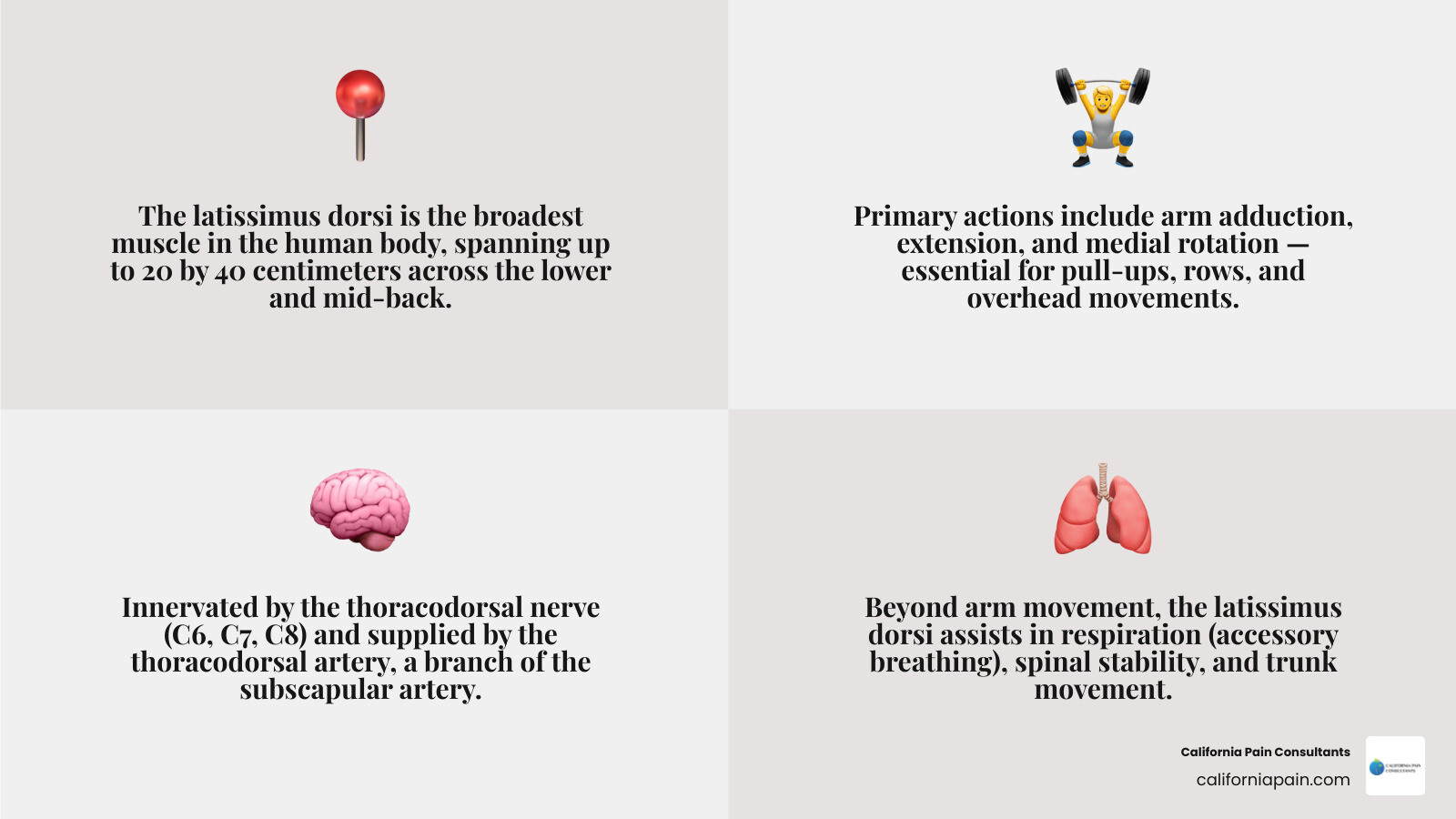
The latissimus dorsi muscle is the broadest muscle in the human body — a large, flat, triangular sheet of tissue that covers most of your lower and mid-back. Here is a quick overview before we dive deeper:
What you need to know at a glance:
| Feature | Detail |
|---|---|
| Location | Lower and mid-back, spanning from spine to upper arm |
| Size | Up to 20 by 40 centimeters |
| Primary actions | Arm adduction, extension, and medial rotation |
| Nerve supply | Thoracodorsal nerve (C6, C7, C8) |
| Blood supply | Thoracodorsal artery (branch of the subscapular artery) |
| Also helps with | Breathing, spinal stability, and trunk movement |
Most people only think about their “lats” when they are doing pull-ups or rows at the gym. But this muscle does far more than move your arms. It connects your spine, pelvis, ribs, and shoulder blade to your upper arm bone (humerus). That means tightness or injury here can quietly cause chronic lower back pain, restricted shoulder movement, and even affect how you breathe.
If you are dealing with unexplained mid-back or shoulder pain that has not responded to typical treatments, the latissimus dorsi is often overlooked — and it may be exactly where to start looking.
I am Dr. Zach Cohen, double board-certified in Anesthesiology and Chronic Pain, and throughout my fellowship training and clinical practice at California Pain Consultants, I have frequently treated patients whose latissimus dorsi muscle dysfunction was the hidden driver behind their persistent back and shoulder pain. In the sections ahead, I will walk you through everything you need to understand about this remarkable muscle — from its anatomy and biomechanics to injury, treatment, and recovery.
Similar topics to latissimus dorsi muscle:
Anatomy of the Latissimus Dorsi Muscle
To appreciate how the latissimus dorsi muscle functions, we first have to look at its sheer scale and structural layout. True to its Latin name, which translates to “the broadest muscle of the back,” it is a structural marvel. While it can measure up to an impressive 20 by 40 centimeters in surface area, it is surprisingly thin, typically measuring less than 1 centimeter in thickness in the average person.
From an evolutionary standpoint, the latissimus dorsi is classified as an “extrinsic” back muscle. This means that during embryonic development, it does not actually start in your back. Instead, it begins its journey within the early arm buds before migrating across the posterior trunk. In fact, an embryo must reach at least 20 mm in length before these migrating muscle fibers finally stretch down to reach the iliac crest. To learn more about how this development fits into the broader muscular system of the back, you can explore the Anatomy, Back, Latissimus Dorsi – StatPearls – NCBI Bookshelf.
Origin and Insertion of the Latissimus Dorsi Muscle
One of the most fascinating aspects of this muscle is how its broad, fan-like origin converges into a single, narrow, twisted point of attachment on the arm. The muscle is divided into four distinct anatomical parts based on where its fibers originate:
- Vertebral part: Attaches to the spinous processes of the lower thoracic vertebrae (T7 through T12) and the lumbar vertebrae.
- Iliac part: Originates from the posterior third of the iliac crest (the top ridge of your hip bone).
- Costal part: Attaches to the lower three or four ribs (9th through 12th ribs).
- Scapular part: Connects to the inferior angle of the scapula (shoulder blade).
Interestingly, the scapular attachment is highly variable. In a detailed study of 100 cadavers, researchers found that 43% of subjects had a substantial amount of muscle fibers directly originating from the scapula, 36% had only a soft fibrous link, and 21% had little to no connecting tissue at all.
All of these broad fiber tracts sweep upward and outward toward the armpit, where they converge. As they approach their destination, the fibers do something unusual: they twist. The superior fibers (from the mid-back) actually insert lower down on the arm, while the inferior fibers (from the lower back and pelvis) spiral to insert higher up.
They end in a flat, quadrilateral tendon about 7 cm long that inserts directly into the floor of the intertubercular (bicipital) groove of the humerus.
Medical students often use a classic, slightly humorous mnemonic to remember this exact relationship: “A Miss Between Two Majors.” In this memory aid, the “Miss” (or “Lady”) represents the Latissimus dorsi, which inserts directly between the Pectoralis major and the Teres major (the “Two Majors”) on the humerus. For a deeper interactive look at these structural connections, check out Latissimus dorsi: Origin, insertion, innervation,function | Kenhub.
| Muscle Region | Origin Points | Insertion Point |
|---|---|---|
| Vertebral | Spinous processes of T7–T12 & Lumbar vertebrae | Floor of the intertubercular groove of the humerus |
| Iliac | Posterior third of the iliac crest | Floor of the intertubercular groove of the humerus |
| Costal | Lower 3 to 4 ribs (9th–12th) | Floor of the intertubercular groove of the humerus |
| Scapular | Inferior angle of the scapula (present in ~79% of people) | Floor of the intertubercular groove of the humerus |
Innervation and Blood Supply
Because the latissimus dorsi muscle is so massive, it requires a highly reliable nerve and vascular network to keep it fueled and coordinated.
It is innervated by the thoracodorsal nerve, which arises from the posterior cord of the brachial plexus, drawing from the C6, C7, and C8 spinal nerve roots. Because of this specific pathway, injuries or disc herniations in the lower cervical spine (particularly C7 lesions) can directly impair latissimus dorsi function, leading to weakness when you try to pull things toward you.
The primary blood supply to the muscle is delivered via the thoracodorsal artery, which is a direct continuation of the subscapular artery. The subscapular artery typically measures between 2 to 5 millimeters in diameter, while the thoracodorsal artery ranges from 1 to 3 millimeters.
This main artery and its accompanying veins (venae comitantes) enter the deep surface of the muscle at a single, highly consistent point: approximately 6 cm to 12 cm from the bifurcation of the subscapular artery, and about 1 cm to 4 cm medial to the muscle’s lateral border.
To prevent tissue death if the main artery is compressed, the muscle also enjoys a backup system of collateral blood flow. This includes branches from the 9th through 11th posterior intercostal arteries and the 1st through 3rd lumbar arteries. Finally, the lymphatic drainage of this massive muscle flows directly into a posterior group of 6 to 7 axillary lymph nodes in the armpit. To explore these vascular pathways in detail, refer to the Latissimus dorsi muscle – e-Anatomy – IMAIOS.
Biomechanics, Respiration, and Trunk Movement

Because the latissimus dorsi muscle crosses several major joints, it is one of only four muscles in the body responsible for true “thoracobrachial motion” — coordinating movements between the spine, pelvis, and upper extremities.
When your spine and pelvis are held in a fixed position, the lats act directly on your shoulder (glenohumeral) joint to perform three primary movements:
- Adduction: Pulling your arm down and in toward the side of your body (like flapping your wings).
- Extension: Moving your arm straight backward behind your body.
- Medial (Internal) Rotation: Rolling your arm inward toward your chest.
However, microscopic structural research shows that this muscle is not just a single, uniform sheet. It actually consists of six distinct groups of muscle fibers that can be independently coordinated by your central nervous system.
Biomechanical data reveals that the average physiological cross-sectional area (PCSA) of the human latissimus dorsi is 5.6 ± 0.5 cm², with an average normalized muscle fiber (fascicle) length of 26.4 ± 1.0 cm. This long fiber length gives the lats an exceptionally high “excursion potential,” meaning they are built to stretch and produce force over a very wide range of motion.
Interestingly, the lumbar-pelvis-costal (LPC) region of the muscle makes up 64% of its total force-generating capacity (PCSA of 3.6 ± 0.3 cm²), while the upper thoracic region accounts for only 36% (2.0 ± 0.2 cm²). This makes the lower portion of your lats the true powerhouse of the muscle. For a detailed architectural analysis of these fiber dynamics, you can read the study published in Architectural analysis and predicted functional capability of the human latissimus dorsi muscle – PMC.
Role of the Latissimus Dorsi Muscle in Respiration and Trunk Movement
While moving the arm is its day job, the latissimus dorsi has a highly active “side hustle” as an accessory muscle of respiration. Because of its attachments to the lower ribs, it acts as the body’s natural “coughing muscle.” During deep inspiration, it helps elevate the lower ribs to expand the chest cavity. During forceful respiratory actions — like a sudden sneeze, a violent coughing fit, or deep, labored breathing — it contracts powerfully to compress the lower rib cage and help force air out of the lungs.
Furthermore, when your arms are fixed in place (for example, when you grip a pull-up bar, use a walker, or walk with crutches), the biomechanics reverse. Instead of the back moving the arm, the lats pull the trunk of your body upward and forward toward your arms.
This function is incredibly important for individuals with paraplegia. By using their lats to stabilize their upper body against parallel bars or crutches, they can actively lift, swing, and control the movement of their pelvis and lower trunk. To explore these full-body movement patterns, check out The Mighty Lats: A Comprehensive Guide to Your Latissimus Dorsi Muscles.
Clinical Significance: Pain, Injury, and Dysfunction
At California Pain Consultants, we frequently see how tightness or weakness in the latissimus dorsi muscle can trigger a domino effect of pain throughout the body. Because it anchors to both the lower spine and the humerus, a short, tight latissimus dorsi will severely restrict your shoulder’s range of motion, particularly during abduction (lifting your arm out to the side) and flexion (reaching overhead).
If you have tight lats and try to reach for a high shelf, your shoulder joint will hit a physical roadblock. To compensate, your body will force your lower back to arch excessively and rotate. Over time, this repetitive, unnatural arching strains the surrounding tissues, leading to chronic lower back, mid-back, and shoulder pain.
Additionally, because the lats are exceptionally powerful internal rotators of the shoulder, they can play a destructive role during extreme physical events. In cases of severe seizures or high-voltage electrical shocks, the violent, involuntary contractions of the lats (alongside the pectoralis major and subscapularis) can completely overpower the shoulder’s weaker external rotators. This overwhelming internal force can actually pull the head of the humerus straight out of its socket, causing a posterior shoulder dislocation. You can read more about how these back muscles interact in our A Comprehensive Guide to Back Muscles.
Latissimus Dorsi Tears vs. Strains
It is crucial to differentiate between a minor latissimus dorsi strain and a severe, structural tear:
- Muscle Strains: These are minor overstretches or microscopic micro-tears within the muscle fibers. They usually develop gradually due to poor posture, desk sitting, or repetitive overhead movements in sports like swimming, golfing, or rowing. Symptoms typically present as a dull, aching tightness or localized stiffness in the mid-back.
- Muscle or Tendon Tears: These are partial or complete ruptures of the muscle belly or its inserting tendon on the arm. They are relatively rare in the general public but occur disproportionately in elite overhead athletes, particularly professional baseball pitchers.
In baseball pitchers, these injuries almost always occur during the high-velocity deceleration phase of a pitch, as the lats contract violently to slow down the rapidly moving arm. Pitchers will often feel a sudden, sharp “pop” or tearing sensation in the back of their armpit, immediately followed by severe pain, swelling, and deep bruising. To learn more about the exercises and movements that can stress or strengthen this area, see our Detailed Guide to Latissimus Dorsi Exercises.
Diagnostic Methods and Imaging
If we suspect a latissimus dorsi injury at one of our California Pain Consultants clinics, we begin with a thorough physical examination. We will often perform a targeted muscle test in a prone position: with the patient lying face down, we ask them to adduct and fully extend their arm, and we then apply downward resistance to the forearm to evaluate strength and pain levels. We also carefully watch for any compensatory spinal arching or pelvic tilting when the patient reaches overhead.
To confirm a diagnosis and rule out other shoulder injuries (such as a rotator cuff tear or a teres major injury), advanced imaging is essential:
- Magnetic Resonance Imaging (MRI): This is the gold standard. An MRI of the shoulder girdle and posterior chest wall provides highly detailed, clear images of the latissimus tendon, allowing us to easily distinguish between a strain, a partial tear, or a complete tendon avulsion from the bone.
- Musculoskeletal Ultrasound: This can be used in the clinic as a rapid, dynamic imaging tool to visualize the muscle fibers and blood flow in real-time.
Treatment, Rehabilitation, and Surgical Applications
For the vast majority of patients suffering from latissimus dorsi pain, conservative, non-surgical treatment is highly effective. However, the ideal recovery pathway depends entirely on the severity of the injury. For a closer look at general rehabilitation and back conditioning, you can read about Back Workouts.
Non-Surgical Pain Management and Physical Therapy
At California Pain Consultants, our board-certified specialists offer a wide range of personalized, non-surgical interventions to relieve latissimus dorsi pain and restore your mobility:
- Conservative Care: During the acute phase of a strain or injury, we recommend resting the area, applying ice packs to reduce swelling, and using over-the-counter anti-inflammatory medications (NSAIDs). Once the acute inflammation subsides, applying heat therapy can help relax the tight muscle fibers and boost local blood flow.
- Trigger Point Injections (TPIs): If chronic stress or poor posture has caused painful, stubborn knots (myofascial trigger points) to form in your lats, we can perform targeted trigger point injections. This highly effective in-office treatment quickly relaxes the muscle, breaks the pain cycle, and provides immediate relief.
- Physical Therapy & Myofascial Release: We work closely with physical therapists to design customized stretching and strengthening programs. This includes hands-on deep tissue massage, myofascial release, and progressive exercises to restore smooth, natural shoulder and spinal mechanics.
For athletes recovering from a partial tear without surgery, physical therapy is a gradual process. However, if an elite athlete undergoes surgical repair for a complete tendon avulsion, the recovery timeline is much longer:
Surgical Applications and Anatomical Variations
Because the latissimus dorsi muscle is so large and has such a reliable, long blood vessel network (with a surgical pedicle length of up to 15 centimeters), it is widely considered the premier “workhorse flap” in reconstructive plastic surgery.
Surgeons frequently use a pedicled latissimus dorsi myocutaneous flap to reconstruct breast tissue following a mastectomy, or to repair massive chest wall defects (such as those seen in Poland’s syndrome). In some advanced cases, surgeons have even wrapped the latissimus dorsi muscle around a failing heart in a procedure called “cardiomyoplasty” to help boost low cardiac output.
Remarkably, because the surrounding back muscles (like the trapezius, rhomboids, and teres major) are highly adaptable, most patients experience surprisingly minimal permanent loss of normal arm function after their latissimus dorsi is harvested.
However, we must always evaluate a patient’s lifestyle thoroughly before surgery; if they rely heavily on crutches or a wheelchair for daily mobility, losing the lats can significantly impact their ability to transfer and move.
Furthermore, donor-site complications are common. Because a large empty space is left behind after the muscle is harvested, fluid collections called seromas frequently form. To manage this, surgeons must keep suction drains in place for at least two weeks or longer post-surgery.
Anatomical Variations to Keep in Mind
When performing surgeries or diagnosing pain in the armpit (axilla) region, we must also account for common anatomical variations:
- The Axillary Arch (Langer’s Muscle): Present in about 7% of the population, this is an anomalous slip of muscle (typically 7 to 10 cm long) that stretches from the latissimus dorsi across the axilla to insert into the humerus or pectoralis major. This extra band of muscle can compress the local nerves and blood vessels, causing thoracic outlet-like symptoms, and can easily confuse surgeons operating in the armpit.
- Dorsoepitrochlearis Brachii (Latissimocondyloideus): Found in approximately 5% of humans, this is an evolutionary remnant of a muscle found in apes. It is a muscular slip that runs from the latissimus dorsi tendon all the way down to the long head of the triceps or the elbow.
To learn more about these fascinating anatomical variations and their surgical implications, you can read the Latissimus dorsi muscle – Wikipedia.
Frequently Asked Questions about the Latissimus Dorsi
How does a tight latissimus dorsi contribute to lower back pain?
Because the latissimus dorsi has massive structural attachments to your lower spine, sacrum, and pelvis via the thick thoracolumbar fascia, any chronic tightness in the muscle acts like a tight structural cable pulling upward on your hips.
This constant upward pull rotates your pelvis forward (anterior pelvic tilt) and forces your lower back into an exaggerated arch (lumbar lordosis). This altered alignment places immense, repetitive stress on the facet joints of your lumbar spine and strains the surrounding lower back muscles, leading to chronic, persistent lower back pain.
Can you function normally if the latissimus dorsi is removed for reconstructive surgery?
Yes, the vast majority of people can return to a completely normal, active life without their latissimus dorsi. This is because your back and shoulders are packed with powerful, highly adaptable synergistic muscles — such as the teres major, trapezius, rhomboids, and posterior deltoids — which quickly step in to take over the work of moving and stabilizing your arm.
The main exception is for individuals who rely heavily on their arms to support their body weight, such as gymnasts, rock climbers, or individuals with paraplegia who depend on crutches or a wheelchair for daily mobility.
What are the best exercises to stretch and strengthen the lats?
To build strong, resilient, and flexible lats, we recommend incorporating a balanced mix of targeted movements into your routine:
- Strengthening: Focus on vertical pulling movements like lat pulldowns and pull-ups, horizontal pulling movements like seated cable rows or bent-over dumbbell rows, and straight-arm movements like cable pullovers to isolate shoulder extension.
- Stretching: The classic “Child’s Pose” from yoga is an excellent way to gently stretch the lats. You can also perform a standing side-bend stretch, reaching one arm up and over your head while holding onto a door frame to deeply stretch the muscle fibers along the side of your back.
Conclusion
The latissimus dorsi muscle is an absolute powerhouse of the human body. As the broadest muscle in your back, it plays a vital role in everything from reaching overhead and stabilizing your spine to helping you breathe and cough. Yet, because of its extensive network of connections, chronic tightness, imbalances, or injuries here can easily manifest as stubborn, unexplained lower back or shoulder pain.
If you are struggling with persistent back pain, restricted shoulder movement, or post-surgical discomfort, you do not have to live with the limitation. At California Pain Consultants, our double board-certified doctors specialize in providing comprehensive, non-surgical pain management tailored to your unique needs.
Serving patients across San Diego, Kearny Mesa, Chula Vista, Rancho Bernardo, La Mesa, and Miramar, California, we are dedicated to delivering compassionate, cutting-edge care to relieve your pain and restore your mobility.
If you are ready to take the first step toward lasting relief, we invite you to contact us today to schedule a personalized evaluation. To learn more about how your back muscles function and how to keep them healthy, visit our comprehensive guide on Back Muscles.