
How to Treat a Pulled Gluteus Medius
Dealing with this in San Diego? Same-day appointments are often available at our 4 clinics.
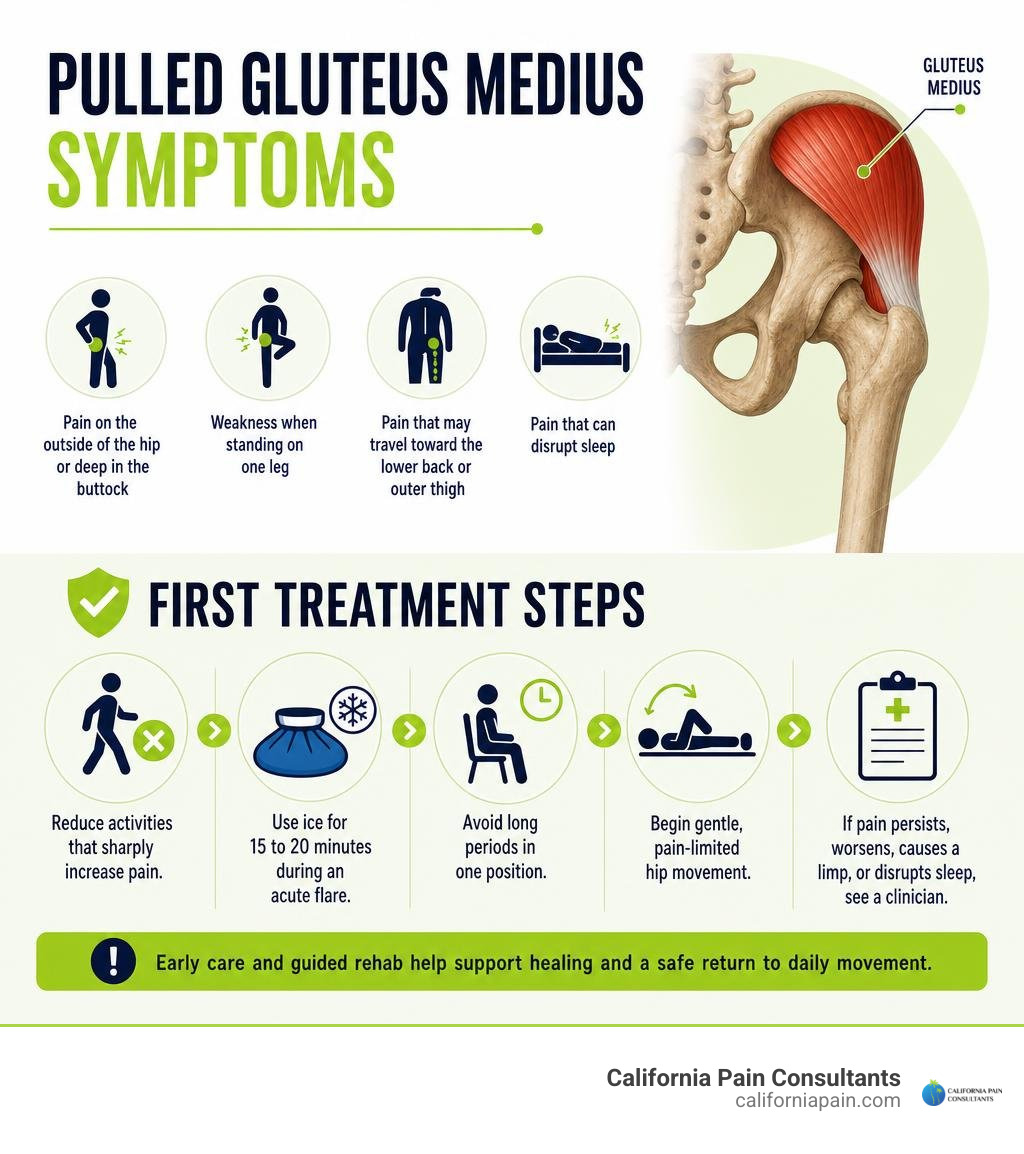
A pulled gluteus medius can cause pain on the outside of the hip or deep in the buttock, weakness when standing on one leg, and pain that may travel toward the lower back or outer thigh. Start by reducing activities that sharply increase pain, using ice for 15 to 20 minutes during an acute flare, avoiding long periods in one position, and beginning gentle, pain-limited hip movement. If pain persists, worsens, causes a limp, or disrupts sleep, see a clinician for an exam and possible imaging.
The gluteus medius is a small but important muscle on the outer side of the pelvis. It helps move your leg outward and keeps your pelvis level with every step. When it is strained, weak, or torn, your body may compensate with a limp, low back pain, or pain that feels like sciatica.
This problem is often mistaken for hip bursitis, spinal stenosis, or a pinched nerve because the symptoms overlap. A clear diagnosis matters, especially for ongoing lateral hip pain or trouble walking.
Dr. Zach Cohen is a double board-certified anesthesiologist and chronic pain physician who helps patients evaluate musculoskeletal pain and pursue personalized, non-surgical treatment when appropriate. His work with chronic hip, back, joint, and nerve pain informs this practical approach to a pulled gluteus medius and safe return to daily movement.
Understanding Pulled Gluteus Medius Symptoms and Causes
The gluteus medius originates along the outer surface of the ilium (the upper blade of the pelvis) and inserts directly onto the lateral facet of the greater trochanter—the bony bump on the outside of your hip. According to clinical anatomical references on the Anatomy, Bony Pelvis and Lower Limb, Gluteus Medius Muscle, this fan-shaped muscle receives its primary nerve supply from the superior gluteal nerve (roots L4, L5, and S1) and is nourished by multiple perforating branches of the deep superior gluteal artery.
When you sustain an injury to this area, the symptoms can be surprisingly widespread. A classic strain often presents as localized lateral hip tenderness, deep buttock aching, and severe single-leg stance instability. Patients frequently report that simple everyday tasks—like stepping out of a low car, stepping sideways while unloading groceries, or climbing a flight of stairs—trigger sharp discomfort.
Another hallmark symptom is nocturnal hip aching and side-sleeping intolerance. Lying on the affected side compresses the irritated muscle tendon against the greater trochanter, while lying on the opposite side allows the top leg to drop forward into adduction, placing the injured tendon under painful mechanical stretch.
Demographic data reveals notable vulnerabilities for this condition. Women are approximately 10 times more likely to suffer a gluteal tendon tear than men, with tears most commonly diagnosed in women between the ages of 50 and 70. This elevated risk is driven by a broader pelvic morphology (which increases the lateral angle and mechanical shear forces placed on the abductor tendons) alongside the systemic decline of circulating estrogen during menopause. Estrogen plays a vital role in maintaining collagen synthesis and tendon elasticity; its reduction can accelerate soft-tissue degeneration, leaving the outer hip vulnerable to a pulled muscle lower back and buttock.
Classic Signs of a Pulled Gluteus Medius vs. Sciatica
Because the gluteus medius sits directly adjacent to major neural pathways in the pelvis, its pain patterns are frequently confused with true lumbar radiculopathy (sciatica). However, clear physical examination markers can help tell them apart:
- The Trendelenburg Sign: When standing solely on the injured leg, a healthy gluteus medius contracts to keep the pelvis level. If the muscle is torn or strained, it cannot support your body weight, causing the pelvis to drop downward on the unsupported side.
- Resisted Hip Abduction Weakness: Lying on your unaffected side and attempting to raise your top leg upward against resistance will reproduce localized pain and reveal marked muscle weakness compared to the opposite side.
- Pain Localization: Sciatica typically creates a sharp, electric, shooting sensation that travels past the knee down into the calf or foot, accompanied by numbness or tingling. In contrast, a gluteus medius injury generates a deep, dull ache over the lateral hip and upper buttock that may refer down the outer thigh toward the knee, but rarely crosses below the joint line, unlike a classic pulled muscle in back.
Common Causes: From Sudden Movement to Degenerative Tendinopathy
Gluteus medius injuries generally fall into two distinct categories:
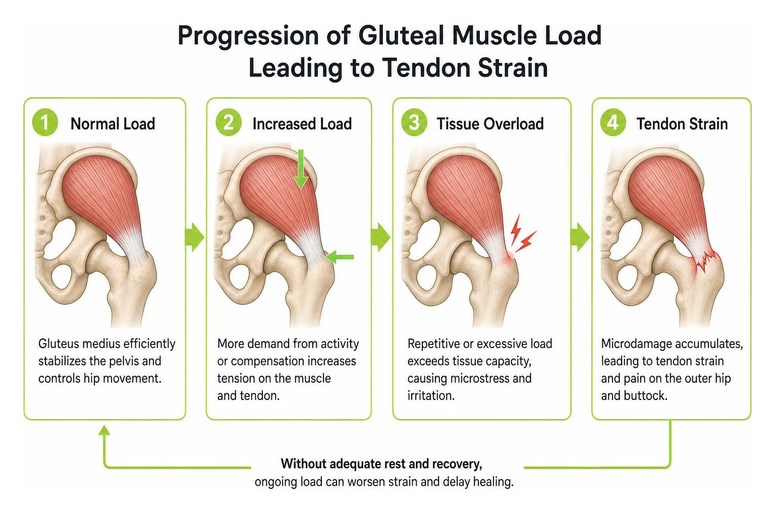
- Acute Mechanical Overload: Sudden, forceful movements such as quick lateral cuts on the tennis court, slips on slick pavement, or an unexpected direct impact from a fall directly onto the greater trochanter can stretch or tear muscle fibers beyond their tensile limits.
- Chronic Microtrauma and Degeneration: The gluteus medius and minimus tendons are widely referred to as the “rotator cuff of the hip.” Just like the rotator cuff in the shoulder, these tendons operate under constant tension during every step of daily life. Over time, repetitive overloading—such as sudden spikes in running mileage, prolonged sedentary sitting without positional variation, or chronic pelvic misalignment—can lead to microtears, collagen breakdown, and degenerative tendinopathy.
Why Gluteus Medius Tears Are Frequently Misdiagnosed
Despite its essential role in human walking, gluteal pathology remains one of the most overlooked causes of lateral hip and pelvic pain in modern medicine. A 2003 survey of orthopedic surgeons revealed that 45 percent of respondents did not know gluteus medius tears could occur. Consequently, patients often spend months or years undergoing treatments for spinal stenosis, sacroiliac joint dysfunction, or trochanteric bursitis without finding lasting relief.
In a clinical pilot evaluation conducted through a spine specialty clinic, 70% of people presenting with lumbosacral and leg pain were found to have painful, weak gluteus medius muscles upon dedicated physical examination. Furthermore, research documented in Gluteus Medius Tendon Rupture as a Source for Back, Buttock and Leg Pain demonstrated that 46% of patients suffering from chronic buttock pain lasting between 1 and 5 years had verifiable gluteus medius tendon tears when evaluated with specialized pelvic MRI.
Many patients presenting with deep buttock discomfort are prematurely funneled into piriformis syndrome treatment or undergo repetitive epidural steroid injections for degenerative spinal changes that are actually incidental, asymptomatic findings.
| Diagnostic Category | Primary Pain Location | Key Physical Exam Finding | Gold Standard Imaging Confirmation |
|---|---|---|---|
| Gluteus Medius Strain / Tear | Lateral hip, outer buttock, upper lateral thigh | Positive Trendelenburg sign; pain on resisted hip abduction | Pelvic MRI (high T2 signal at lateral trochanteric facet) or high-resolution ultrasound |
| Trochanteric Bursitis | Localized directly over the greater trochanteric bursa | Extreme pinpoint tenderness over the lateral bone; minimal weakness | Ultrasound showing bursal fluid distension without tendon disruption |
| Lumbar Radiculopathy (Sciatica) | Lower back radiating below the knee to calf/foot | Positive Straight Leg Raise (SLR); dermatomal sensory changes | Lumbar Spine MRI (nerve root compression / herniation) |
Accurate differential diagnosis requires high-resolution magnetic resonance imaging (MRI) or specialized musculoskeletal ultrasound. In an ultrasound study of 75 patients presenting with greater trochanteric pain, 63 were confirmed to have gluteal tendinopathy, with 25 displaying full-thickness or partial-thickness abductor tendon tears.
The 5-Step Treatment Protocol for Lasting Hip Recovery
Healing a pulled or torn gluteus medius does not mean staying in bed for weeks. In our clinical practice, we emphasize an active, progressive rehabilitation framework focused on reducing inflammation, restoring pelvic stability, and rebuilding tendon resilience. For a complete overview of hip rehabilitation strategies, review our hip pain relief complete guide. You can also explore how to relieve muscle strain fast by using simple techniques to manage your symptoms early on.
Step 1: Protect the Tendon and Relieve Acute Inflammation
The first priority during an acute flare is removing excessive compressive and tensile stress from the tendon insertion.
- Contralateral Cane Offloading: If you are limping or experiencing sharp pain during walking, use a single-point cane in the hand opposite your injured hip. Holding the cane on the unaffected side reduces the muscular force your injured gluteus medius must generate to keep your pelvis level by over 50%.
- Targeted Cryotherapy: Apply ice packs wrapped in a thin towel over the lateral hip for 15 to 20 minutes, 3 to 4 times daily, to calm acute localized inflammation.
- Posture Adjustments: Avoid crossing your legs while seated, as this forces the injured tendon to stretch tightly over the greater trochanter. When resting in bed, place a pillow between your knees if sleeping on your uninjured side, or sleep on your back.
- To learn more about initial self-care steps, see how to treat muscle strain even if youve never tried it before.
Step 2: Early Isometric Activation and Gentle Mobility
Complete rest can lead to muscle atrophy and tendon stiffness. As soon as acute pain begins to settle, introduce submaximal isometric muscle contractions to stimulate local blood flow and maintain neuromuscular pathways without aggravating the tendon fibers.
- Standing Lateral Wall Press: Stand tall with your injured side facing a wall, roughly 6 inches away. Bend your knee slightly and gently press the outside of your knee and lower thigh into the wall. Hold this static contraction for 5 to 10 seconds at roughly 30% to 50% of your maximum effort. Repeat for 8 to 10 repetitions.
- Gentle Lumbar and Hip Capsule Mobility: Maintain flexibility throughout your lumbar spine and pelvis by incorporating supported pelvic tilts and gentle supine knee-to-chest movements. For detailed guidance on preserving spinal mobility during hip rehabilitation, see how to stretch lower back.
Step 3: Strengthening Exercises for a Pulled Gluteus Medius
Once isometric exercises are pain-free, progress to active strengthening. Electromyographic (EMG) studies have reshaped how physical therapists approach gluteal rehabilitation. As detailed in the Gluteus Medius Anatomy: Function, Exercises & Muscle Guide, traditional clamshell exercises often activate the anterior tensor fasciae latae (TFL) and hip flexors more than the gluteus medius itself.
To isolate the three distinct segments of the gluteus medius (anterior, middle, and posterior fibers), prioritize the following evidence-based exercises, which are also highlighted in our collection of hip pain relief exercises:
- Side-Lying Hip Abductions: Lie on your unaffected side with your bottom leg bent for stability. Keep your top leg straight, slightly behind the midline of your torso, with your toes pointed slightly downward. Slowly raise the top leg toward the ceiling (no higher than 30 to 40 degrees) and lower it under control. Perform 2 to 3 sets of 10 to 12 repetitions.
- Pelvic Hip Hikes: Stand with the foot of your unaffected leg on a step or small box, allowing your injured leg to hang freely off the side. Keep both knees straight. Lower your free hip down slightly toward the floor, then use the gluteus medius of your stance leg to hike the opposite hip upward above level. Perform 2 to 3 sets of 10 repetitions.
- Single-Leg Supported Split Squats: Place one foot forward and one foot back in a staggered stance. Slowly lower your back knee toward the floor while ensuring your front knee stays aligned directly over your second toe without collapsing inward. Perform 2 to 3 sets of 8 to 10 repetitions per side.
Step 4: Targeted Non-Surgical Pain Therapies
When severe gluteal tendinopathy or partial tearing does not respond to exercise therapy alone, advanced interventional pain management can help support tissue healing:
- Platelet-Rich Plasma (PRP) Injections: Autologous PRP therapy involves concentrating growth factors from your own blood and injecting them directly into the degenerated tendon under ultrasound guidance to support tissue remodeling.
- Ultrasound-Guided Trochanteric Injections: When severe secondary trochanteric bursitis limits your ability to participate in physical therapy, a precisely targeted bursal injection can provide substantial pain relief.
- Percutaneous Ultrasound Tenotomy: A minimally invasive procedure where an ultrasound-guided micro-tip breaks down and removes localized calcifications and scarred tendon tissue to encourage healthy collagen regeneration.
- Integrating these treatments alongside specialized back pain physical therapy helps address compensatory muscle imbalances across the spine and pelvis.
Step 5: Dynamic Gait Retraining and Long-Term Prevention
The final stage of rehabilitation focuses on functional movement patterns to prevent re-injury:
- Lateral Band Walks: Place a light resistance band around your ankles or just above your knees. Assume a quarter-squat position with your feet shoulder-width apart. Take controlled steps laterally, keeping your knees pushed outward against the band tension and your torso upright.
- Single-Leg Balance Progressions: Practice balancing on your injured leg for 30 seconds at a time, progressing from a stable flat floor to an unstable balance pad or foam cushion.
- Kinetic Chain Alignment: Ensure your running and walking mechanics avoid excessive hip drop, dynamic knee valgus (knees caving inward), or over-striding. For a broader look at kinetic chain health, review our lower back pain solutions ultimate guide.
When Conservative Care Fails: Advanced Interventions and Surgical Repair
The vast majority of gluteus medius strains and partial tears heal successfully with structured conservative care. However, when full-thickness tears, significant muscle retraction, or chronic severe pain persist despite months of dedicated non-surgical therapy, surgical evaluation may be warranted.
Modern surgical interventions have advanced significantly:
- Minimally Invasive Endoscopic Repair: As detailed in Endoscopic Repair of Partial Gluteus Medius Tear: Staple Configuration With Suture Anchors, orthopedic surgeons can use small arthroscopic incisions and suture anchors arranged in a staple configuration to reattach torn abductor tendons directly onto the greater trochanter. This technique often incorporates bioinductive collagen patches to encourage biological tissue integration while minimizing surgical trauma.
- Muscle Flap Transfer and Synthetic Augmentation: For chronic, massive, or retracted tears where the tendon tissue has experienced fatty degeneration, primary repair alone may carry a high failure rate. In these complex cases, techniques such as Gluteus Medius Repair using a Gluteus Maximus Flap Augmented with Synthetic Polyethylene Mesh: Surgical Technique and Case Series can be considered. This procedure mobilizes a portion of the anterior gluteus maximus muscle and reinforces it with a synthetic polyethylene terephthalate mesh to restore hip abduction mechanics and resolve severe Trendelenburg gait.
Post-operative recovery following surgical tendon repair typically requires strict non-weight-bearing precautions with crutches and an abduction brace for approximately six weeks, followed by several months of progressive physical therapy.
Frequently Asked Questions About Gluteus Medius Strains
How long does it take for a strained gluteus medius to heal?
Recovery timelines depend on the structural severity of the strain and your adherence to progressive loading:
- Grade 1 (Mild Strain): Microscopic fiber damage without structural discontinuity typically resolves in 2 to 4 weeks with relative rest and gentle mobility.
- Grade 2 (Moderate Partial Tear): Significant fiber disruption with marked weakness generally requires 6 to 12 weeks of structured physical therapy.
- Grade 3 (Full-Thickness Tear / Tendon Avulsion): Severe tears may require 3 to 6 months or longer of intensive conservative rehabilitation or surgical intervention.
- To learn more about healing timelines across different gluteal injuries, read our guide on pulled muscle in buttock how long to heal.
Can a weak or torn gluteus medius cause lower back and knee pain?
Yes. The gluteus medius serves as the primary lateral stabilizer of the pelvis. When this muscle is weak or painful, the pelvis drops downward every time you take a step. To keep you upright, your lumbar spine compensates by bending sideways, placing uneven mechanical strain on your facet joints, intervertebral discs, and the quadratus lumborum muscle.
Down the kinetic chain, gluteal weakness allows the femur to rotate inward, leading to patellofemoral tracking issues, increased IT band tension, and elevated stress on the anterior cruciate ligament (ACL). Incorporating targeted exercises for lower back pain alongside hip rehabilitation helps protect the entire lower body.
Is walking good for gluteus medius pain?
Walking is beneficial when performed correctly, but it must be managed carefully during recovery. Extended walks that cause a visible limp or trigger sharp lateral hip pain can worsen tendon inflammation.
Focus on short, frequent walks on flat surfaces rather than long, fatiguing sessions. Shortening your stride slightly and maintaining a moderate cadence can decrease the lateral peak forces applied to the greater trochanter. For additional strategies on maintaining an active lifestyle while recovering from musculoskeletal discomfort, explore our guide to natural back pain relief.
Conclusion
A pulled gluteus medius can be a frustrating and commonly misdiagnosed cause of chronic hip, buttock, and lower back discomfort. Because its pain referral patterns mimic sciatica and lumbar spinal stenosis, obtaining an accurate diagnosis through physical examination and imaging is essential.
By following a structured, step-by-step recovery plan—protecting the tendon during acute flare-ups, reintroducing gentle isometric contractions, and progressively building abductor strength—most individuals can resolve their symptoms and return to comfortable movement without invasive surgery.
At California Pain Consultants, our board-certified physicians specialize in comprehensive, non-surgical pain management across our clinic locations in San Diego, Kearny Mesa, Chula Vista, Rancho Bernardo, La Mesa, and Miramar, California. If you are struggling with persistent lateral hip pain, an undiagnosed limp, or discomfort that disrupts your sleep, contact our team today to schedule an evaluation. For further information on recovery expectations, explore our resource on pulled muscle in buttock how long to heal.